




Breast ultrasound and mammography both help to check for abnormalities in the breast, but they are not the same method and they do not fully replace each other.
Put simply:
Breast ultrasound → uses sound waves to look at the structures inside the breast, and is particularly useful when an abnormal lump needs to be assessed.
Mammography → uses low-dose X-rays to detect changes in breast tissue, including some very small lesions that cannot yet be felt.
How do breast ultrasound and mammography differ?
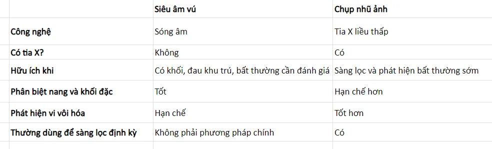
The important point is that each method sees different features of the breast tissue.
That is why doctors sometimes order both rather than choosing one or the other.
When should you have a breast ultrasound?
1. When you can feel a lump in the breast
This is one of the most common reasons for an ultrasound.
An ultrasound can help to determine whether the lump is a fluid-filled cyst or a solid lump, which helps the doctor decide whether further monitoring or a biopsy is needed.
2. When there is pain or a localised change in the breast
Your doctor may consider an ultrasound if you have:
- A newly appeared lump
- Pain in one specific spot
- Abnormal nipple discharge
- Changes in the skin or in the structure of the breast
- An abnormal area found during an examination
This is an assessment of symptoms, which is different from routine screening carried out when you have no symptoms at all.
3. Young people with a breast lump
In people under 30 with a new breast lump, ultrasound is usually the first imaging method used.
This does not mean that young women never need mammography. If the ultrasound finds a suspicious lesion, the doctor may still order further investigations.
4. When breast tissue is dense
Dense breast tissue can make some lesions hard to see on a mammogram, because both dense tissue and some abnormalities appear white.
In some cases, the doctor may consider a supplementary ultrasound after the mammogram.
However, dense breast tissue does not always mean an additional ultrasound is required. The evidence on the benefit of supplementary ultrasound for screening every woman with dense breast tissue is still not entirely clear.
When should you have a mammogram?
1. When you reach the age for breast cancer screening
Mammography is the main imaging method used to screen for breast cancer in women without symptoms.
Professional bodies may recommend slightly different screening schedules.
For example, the USPSTF currently recommends that women at average risk aged 40–74 have a mammogram every 2 years. Meanwhile, some other specialist organisations recommend starting at 40 with a more frequent schedule.
So do not simply remember a single age. The right schedule also depends on your individual risk and the guidance your healthcare provider follows.
2. When the doctor needs to assess an abnormality
Beyond routine screening, diagnostic mammography may be carried out when there is:
- A lump in the breast
- Nipple discharge
- A change in the shape or size of the breast
- An abnormality on a previous scan
For women from about 30 onwards with a new lump, the doctor can often use diagnostic mammography combined with ultrasound for a more complete assessment.
Why can mammography see things that ultrasound may miss?
One very important point is microcalcifications.
These are very tiny specks of calcium in the breast tissue. Most are not cancer, but certain patterns of microcalcification can be linked to pre-cancerous changes or early-stage cancer.
Mammography is better than ultrasound at detecting these changes.
That is why:
A “normal ultrasound” does not mean you can skip mammography once you have reached screening age or belong to a group that needs screening.
Do you need both an ultrasound and a mammogram?
Not everyone does.
For example:
A young person under 30 with a new lump
→ Ultrasound is usually preferred first.
A woman of screening age with no symptoms
→ Mammography is usually the main method.
An abnormal lump or an unclear mammogram result
→ An additional ultrasound may be needed.
Dense breast tissue or high risk
→ The doctor may consider an additional ultrasound or MRI depending on the case.
So do not choose a test based only on the idea that “ultrasound has no X-rays, so it is better”.
Not using X-rays is an advantage of ultrasound, but that does not make it a complete replacement for mammography.
When should you not wait for your screening appointment?
You should see a doctor if you notice:
- A new lump in the breast or armpit
- Abnormal nipple discharge, especially if bloodstained
- A nipple that has newly become inverted
- Dimpling, thickening or any unusual change in the breast skin
- A clear change in the shape or size of one breast
- Persistent localised pain or a new abnormality
Most breast lumps are benign, but a new lump still needs to be assessed rather than monitored on your own for a long period.
Do not decide on age alone
Age is an important factor but not the only one.
Doctors also consider:
- A family history of breast cancer
- Your personal medical history
- Breast tissue density
- High-risk gene mutations such as BRCA1/BRCA2
- A history of radiotherapy to the chest
- Previous mammogram or biopsy results
People at high risk may need a different screening programme from those at average risk, including breast MRI in some cases.
Book a breast health check on Halza
Through Halza, you can browse and book breast examinations, breast ultrasound, mammography and breast cancer screening at partner hospitals and clinics.
Keeping the results of previous checks also makes it easier for doctors to compare changes in breast tissue over time.
Ultrasound helps to show the features of a lump clearly. Mammography helps to detect changes that you may not yet be able to feel. What matters is not choosing whichever method is “better”, but choosing the right method for the right situation.
The information in this article is for reference only and does not replace the advice, diagnosis or instructions of a doctor. Breast cancer screening methods and schedules should be tailored to each person's age and risk.
Frequently asked questions
Can a breast ultrasound detect breast cancer?
It can detect some suspicious lesions, but ultrasound is not a breast cancer screening method that fully replaces mammography.
Ultrasound is particularly useful for seeing whether a lump is:
- A fluid-filled cyst
- A solid lump
- Or a lesion needing further assessment
Some cancers, especially lesions that show up as microcalcifications, can be seen on a mammogram but are not clear on ultrasound.
Is a mammogram painful?
During the scan, the breast is compressed between two plates of the machine for a few seconds to produce a clearer image.
You may feel tightness or discomfort, especially if your breasts are tender before your period. The sensation usually lasts only a short time.
Does mammography use X-rays?
Yes.
Mammography uses low-dose X-rays to produce images of the breast tissue. It is a technique that is widely used in breast cancer screening.
Ultrasound, on the other hand, does not use X-rays.
References
- RadiologyInfo – Breast Ultrasound
- RadiologyInfo – Mammography
- RadiologyInfo – Palpable Breast Masses
- National Cancer Institute – Breast Cancer Screening & Dense Breasts
- USPSTF – Breast Cancer Screening Recommendation, 2024
 Back to all articles


